Vol.75, No.1 January 2013
Explaination of the cover photograph
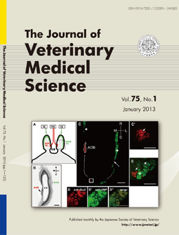
Existence of Gαi2-Expressing Axon Terminals in the Goat Main Olfactory Bulb
Hiromi Ohara, et al. (pp. 85–88)
(A): A horizontal view of the OB (B) is schematically illustrated. The axonal projections of the sensory neurons in the olfactory epithelium (OE) and the vomeronasal organ (VNO) to the glomerular layer (GL) in the main olfactory bulb (MOB; green area) and accessory olfactory bulb (AOB; red area) were shown. (C): OB sections were labeled with anti-Gαi2 (red) and anti-Gαolf (green). The GL of the MOB expressed Gαolf (asterisks), whereas the GL of the AOB expressed Gαi2p (red area enclosed by dotted line). In addition to the AOB, small Gαi2-immunoreactive clusters located outside the AOB (boxed area) were identified.
High magnification of these clusters (C’ and E) showed that they included at least several Gαi2-immunoreactive glomeruli (arrow). (D): OMP was strongly expressed in these clusters, and the expression overlapped with that of Gαi2 (2D–D′′). These results suggest that the axon terminals of the Gαi2-expressing mature sensory neurons in the olfactory organs converged onto the glomeruli in the caudal MOB.
This number is available on J-STAGE
http://www.jstage.jst.go.jp/browse/jvms/75/1/_contents