Vol.77, No.6 June 2015
Explanation of the cover photographs
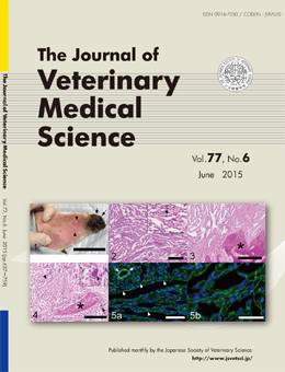
Lymphangiosarcoma with bone formation of the auricle in a dog
Takayuki MINESHIGE et al. (pp. 739–742)
Lymphangiosarcoma of a dog. Gross appearance of the tumor mass (arrows) and two millet-sized daughter nodules (arrowheads) on the auricle (1). Histologic feature of the tumor comprising numerous clefts with no erythrocytes (2). Bone formation (*) in the well-developed stroma separated from neoplastic cells (3). Osteoclast-like giant cells (arrows) and osteoblasts around the bone tissue (*) and osteoid formation (arrowhead). (4). Immunofluorescence showing linear or granular laminin expression around neoplastic vascular channels (5a and 5b).
This number is available on J-STAGE
https://www.jstage.jst.go.jp/browse/jvms/77/6/_contents