Vol.78, No.2 February 2016
Explanation of the cover photographs
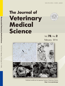
Culture and propagation of microsporidia of veterinary interest
Maria Anete LALLO et al. (pp. 171–176)
The figures depict the cell culture of Encephalitozoon sp., an important Microsporidia for Veterinary Medicine. Rabbit kidney (RK) cells are distended by E. cuniculi spores and other stages, as is observed by inverted microscope (a). By transmission electron microscopy (b) we can observe a parasitophorous vacuole full of sporogonial stages and spores of E. intestinalis in Vero cells (green monkey Kidney cell).
E. cuniculi spores grow inside the cells, and the development forms of the pathogen distend the cells and promote their rupture, allowing the release of spores as seen by scanning electron microscopy (c, d).
This number is available on J-STAGE
https://www.jstage.jst.go.jp/browse/jvms/78/2/_contents