Vol.79, No.8 August 2017
Explanation of the cover photograph
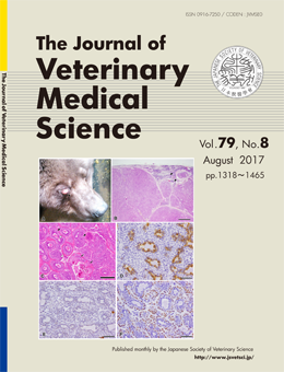
Basal cell adenocarcinoma in the gland of the third eyelid of a brown bear (Ursus arctos)
Hiroki Sakai et al. (pp. 1348-1351)
Grossly, the swollen third eyelid of the right eye was protruded (A). Microscopically, the tumor was compartmentalized with connective tissue, and exhibited mildly infiltrative proliferation, resulting in the involvement of adjunct glands of the third eyelid (B). The tumor consisted of multi-stratified glandular structures of round cells, prominently surrounded by eosinophilic thick basal lamina. The innermost neoplastic cells of the glandular structures contained eosinophilic secretions in the lumen, and there were basaloid neoplastic cells surrounding the laminal epithelial neoplastic cells. A few squamous metaplastic foci were observed (C). The innermost neoplastic cells of the glandular structures were positive for cytokeratin 8/18 (D), and the nuclei of the basaloid neoplastic cells were positive for p63 (F). Both types of neoplastic cells were negative for α-smooth muscle actin (E).
This number is available on J-STAGE
https://www.jstage.jst.go.jp/browse/jvms/79/8/_contents