Vol.80, No.8 August 2018
Explanation of the cover photographs
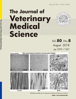
Histopathological and electron microscopic study in dogs with patellar luxation and skin hyperextensibility
Kazunori UEDA et al. (pp. 1309–1316)
Transmission electron microscopic findings of cross (A–C) and longitudinal (D–F) sections of the skin. While the control group (A, D) exhibited thick bundles of closely packed collagen fibrils (mean diameter, 103.67 ± 15.58 nm), dogs with patellar luxation (B, E: medial patellar luxation (MPL); C, F: medial-lateral patellar luxation (MLPL)) exhibited loosely packed bundles of significantly thinner collagen fibrils (mean diameter, 71.51 ± 16.49 nm), with the MLPL samples showing disorganized fibrils and heterogeneity in collagen fibril diameter.
This number is available on J-STAGE
https://www.jstage.jst.go.jp/browse/jvms/80/8/_contents