Vol.80, No.7 July 2018

Explanation of the cover photographs
Surgery for partial atrioventricular septal defect with pulmonary hypertension in an adult dog
Seijirow GOYA et al. (pp. 1183–1189)
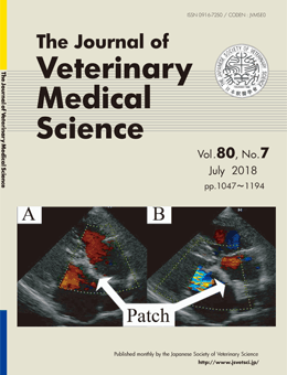
A 4-year-old, 5.9-kg female Japanese Spitz presented with syncope and exercise intolerance. Echocardiography revealed an ostium primum atrial septal defect, a cleft mitral valve, mitral valve regurgitation, and tricuspid regurgitation, leading to a diagnosis of partial atrioventricular septal defect with moderate pulmonary hypertension. Open-heart surgery was performed using cardiopulmonary bypass. This graph shows that color-flow Doppler echocardiogram images at 2 months after the surgery. In the diastolic phase (A), an autologous pericardium patch fixed with glutaraldehyde occluded the defect and left-to-right shunting flow was not observed. In the systolic phase (B), tricuspid regurgitation was absent, but mitral regurgitation persisted.
This number is available on J-STAGE
https://www.jstage.jst.go.jp/browse/jvms/80/7/_contents