Vol.75, No.11 November 2013

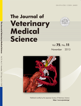
Explanation of the cover photographs
Computed Tomography Angiography of Situs Inversus, Portosystemic Shunt and Multiple Vena Cava Anomalies in a Dog
Heejin OUI et al. (pp. 1525-1528)
Volume-rendered computed tomography reconstruction of the dog with multiple vascular malformations and situs inversus viewed from a left ventral perspective. The portal branch (red) showed a serpentine course and entered into the left hepatic vein (yellow). From this image, extrahepatic single portosystemic shunt was confirmed.
This number is available on J-STAGE
http://www.jstage.jst.go.jp/browse/jvms/75/11/_contents