Vol.76, No.5 May 2014

Explanation of the cover photograph
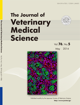
Trichoblastoma with Abundant Plump Stromal Cells in a Dog
Takayuki Mineshige et al. (pp. 735-739)
Double-labeled immunofluorescence microscopy of the tumor tissue. Green color indicates positive staining for CK clone AE1/AE3. Red color shows positive staining for vimentin. Nuclei are colored blue with 4,6-diamino-2-phenylindole. Neoplastic epithelial cells are positive for CK AE1/AE3 but not for vimentin. In contrast, plump stromal cells are positive for vimentin but not for CK AE1/AE3.
This number is available on J-STAGE
https://www.jstage.jst.go.jp/browse/jvms/76/5/_contents