Vol.77, No.11 November 2015

Explanation of the cover photographs
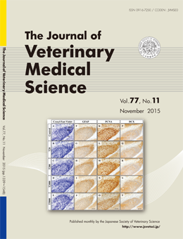
Immunohistochemical analysis of 2,3,7,8-tetrachlorodibenzo-p-dioxin (TCDD) toxicity on the developmental dentate gyrus and hippocampal fimbria in fetal mice
Yoshihiro Kobayashi et al. (pp. 1355- 1361)
Representative histology and immunohistochemistry in the DG in the hippocampus at E18.5. (A–E) The cell density in the DG was decreased in the T5000 compared with the control group, whereas no obvious changes were observed in the shape or somatic size of cells (E). (F–J) The fibrous immunoreactivities of GFAP were detected, but were decreased in the T5000 group (J). (K–O) The numbers of PCNA- positive cells were smaller in the T5000 (O) than the other groups (K–N). (P–T) The intensity of DCX staining did not differ between the control and TCDD-exposed groups.
This number is available on J-STAGE
https://www.jstage.jst.go.jp/browse/jvms/77/11/_contents