Vol.75, No.12 December 2013
Explanation of the cover photographs
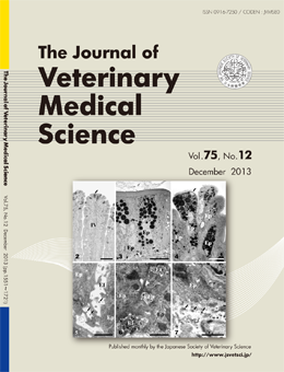
Ultrastructural Demonstration of the Absorption and Transportation of Minute Chylomicrons
by Subepithelial Blood Capillaries in Rat Jejunal Villi
Ei-ichirou TAKAHARA et al. (pp. 1563-1569)
A light microscopic image of lipid droplets producing epithelial cells in the jejunal villi (2). Electron microscopic figures of large and highly electron dense fat droplets in the orderly epithelial cells (3) and the exfoliating epithelial cells (4) of the villous apices.
Electron microscopic figures of highly electron dense chylomicrons in the apical portion (5) and the basal portion (6) of the epithelial intercellular spaces in the villous apices. An electron microscopic figure of highly electron dense chylomicrons in the area between the subepithelial blood capillaries in the villous apex (7). Block staining by 1.0% imidazole-1.0% paraphenylenediamine.
This number is available on J-STAGE
http://www.jstage.jst.go.jp/browse/jvms/75/12/_contents