Vol.77, No.12 December 2015
Explanation of the cover photographs
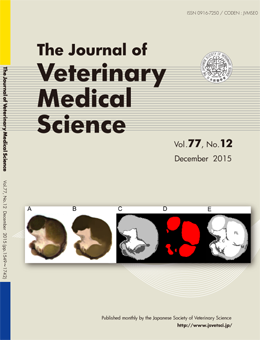
Analysis of the equine ovarian structure during the first twelve months of life by three-dimensional internal structure microscopy
Mamiko ONO et al. (pp. 1599–1603)
Extraction of the cortex and medulla of the equine ovary. A: Original image. B: Regions with similar colors were grouped by the mean shift method. C: The image was divided into three classes, the background, cortex (white) and medulla (gray), by the k-means method. D: Regions corresponding to the follicles (red) were extracted from the original image. E: A composite image of C and D. The follicles detected within the medulla were regrouped with the cortex. FL, follicle; C, cortex; M, medulla.
This number is available on J-STAGE
https://www.jstage.jst.go.jp/browse/jvms/77/12/_contents