Vol.78, No.10 October 2016
Explanation of the cover photographs
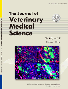
Transmission of atypical scrapie to homozygous ARQ sheep
Hiroyuki OKADA, et al. (pp.1619–1624)
Dual immunofluorescence staining against the disease-associated prion protein (PrPSc; a–d, green) and glial fibrillary acid protein (GFAP) for astrocytes (a and b, red) or ionized calcium-binding adaptor molecule (Iba-1) for microglia (c and d, red). Fine to coarse granular PrPSc deposits can be observed principally in the neuropil and associated with the cytoplasmic protrusions (yellow) of astrocytes (a) and microglia (c). Boxed regions in panels (a) and (c) are shown at higher magnification in panels (b) and (d), respectively. PrPSc deposits can be discerned within astrocytes and microglia or at the periphery of these cells. bv: Blood vessel.
This number is available on J-STAGE
https://www.jstage.jst.go.jp/browse/jvms/78/10/_contents