Vol.79, No.11 November 2017
Explanation of the cover photographs
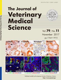
Immunolocalization of β-catenin, E-cadherin and N-cadherin in neonate and adult rat kidney
Naomi TERADA et al. (pp. 1785-1790)
A-D: Immunohistochemistry in renal cortex of neonate kidney on day 1. Expression of β-catenin (A) and E-cadherin (B) are observed in the developing renal tubular epithelial cells (inset). Few blastemal cells react to β-catenin among developing renal tubules (arrows). N-cadherin (C) is weakly expressed in some parts of developing tubules. Inset show positive reaction in renal epithelial cells of higher magnification. D: The developing renal tubules do not express vimentin, but blastemal cell-derived mesenchymal cells strongly express vimentin.
E-G: Double immunofluorescence in renal cortex of neonate kidney on day 1. β-catenin (E, red)- or N-cadherin (F, red)-expressing epithelial cells are corresponding to some E-cadherin (green)-expressing cells in the developing renal tubules (arrowhead, inset). Yellow color indicates double positive reaction. Some blastemal cells show positive reaction with β-catenin among developing renal tubules (arrows). G: Vimentin (red)-expressing blastemal cells are not corresponding to E-cadherin (green)-expressing cells. Inset shows E-cadherin localization in renal epithelial cells of higher magnification.
This number is available on J-STAGE
https://www.jstage.jst.go.jp/browse/jvms/79/11/_contents