Vol.80, No.2 February 2018
Explanation of the cover photographs
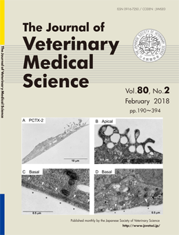
Inhibition of actin polymerization by marine toxin pectenotoxin-2
Masatoshi HORI et al. (pp. 225–234)
Ultrastructure of A7r5 cells treated with PCTX 2
Th. typical ultra-structures of A7r5 smooth muscle cells in the presence of PCTX-2 (100 nM) chosen from images of at least 10 cells are shown here. After 30 min incubation with PCTX-2 at 37℃, the cells were fixed. The open arrowhead and the asterisk in the basal membrane (C and D) show the forming focal adhesion containing actin filaments. The electron-density of actin filaments in the focal adhesion was lower than that in contro1 cells (see Fig. 9 of the text). The apical actin filaments associated with the apical plasma membrane disappeared completely (B). The closed arrowhead in D indicates microtubules. The bar indicates 10 μm in A and 0.5 μm in B, C and D. N: nuclei. M: mitochondria. All pictures were taken by Dr. F. Yazama.
This number is available on J-STAGE
https://www.jstage.jst.go.jp/browse/jvms/80/2/_contents